
X-rays are a form of imaging that is commonly used in dental practice to diagnose bone loss, cavities, and other hidden dental structures. They can also be used to diagnose both benign and malignant masses.
Digital vs. film
Compared to conventional film, digital X-rays are a safer alternative. They do not require chemical development, are easier to store, and provide higher-quality images. They are also less likely to be underexposed. This makes it easier to see problems that may not be noticeable with a conventional X-ray.
Digital imaging is a major change in the dental industry. It allows for better diagnostics and treatment planning. These digital images also allow the clinician to manipulate them in ways that were previously impossible. The latest digital imaging systems also allow for greater diagnostic flexibility, improved image enhancement, and better follow-up conditions.
In the dental field, digital imaging has many advantages over conventional film. It improves patient care by providing high-quality images of bones, teeth, and gums, and allows the clinician to perform more efficient imaging tasks.
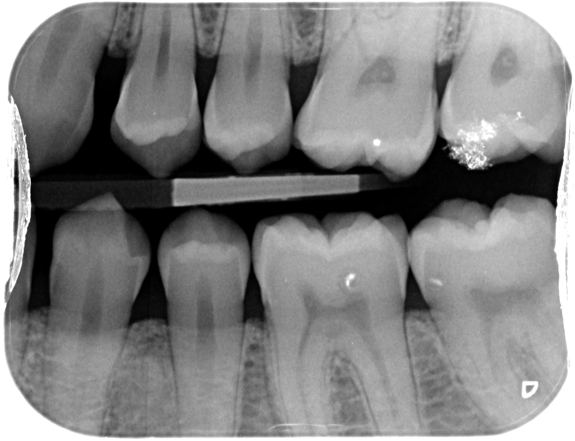
Periapical (limited) X-rays
Compared to conventional radiographs, periapical (limited) X-rays provide a more accurate picture of the crown, roots, and supporting bone of a tooth. They may also reveal changes in the bone or the position of teeth. In addition, a periapical X-ray can determine the depth of decay, whether there are cysts, and whether the tooth is impacted.
In a recent study, a full mouth series of periapical radiographs was compared to cone beam computed tomography (CBCT) scans in patients with apical periodontitis. The study included 101 patients with previous endodontic treatments. They were evaluated by two independent operators. They found that CBCT scans yielded a higher LR+ than SPP.
The study found that periapical films are useful for diagnosing cracked roots. However, the extent of radiographic detection depends on the localization of the lesion in the bone. The lesion was most easily identified when it was near the cortex, but it was not easy to detect lesions in the cancellous bone.
Periapical (limited) technique
Various authors have reported on the diagnostic ability of cone beam computed tomography (CBCT) and its use in dental radiography. These studies were conducted using conventional intraoral and extraoral techniques. Some of the authors did not have experience in digital radiography. Others claimed difficulty in positioning CCD sensors. In the present study, we performed a study using LCBCT.
The cone of the radiographic device was positioned in the opposite direction from the tooth. The cone reached the perpendicular angle of the film. After repositioning the teeth, new radiographs were taken. The tooth was not referred for root canal therapy.
The mean number of periapical lesions detected with periapical films was 0.95. The CBCT system yielded a mean number of 1.55 per tooth. The LCBCT system also showed higher specificity than the SPP system. However, a higher level of interobserver agreement indicated a consistent level of performance for the LCBCT system.
Ethnicity affects the likelihood of LBW
LBW has long been an issue in the United States. A number of clinical and public health measures have been implemented in the hopes of reducing the prevalence of LBW infants. However, the problem persists throughout the country. In particular, LBW has been found to be more prevalent among African American women.
The presence of LBW is linked to both perinatal and genetic factors. Prenatal health behaviors, maternal age and educational level, and genetics all play an important role in determining the likelihood of having a LBW infant. In addition, LBW can result in short-term morbidity and long-term disability such as cerebral palsy and blindness.
Among women, education level is a strong determinant of LBW prevalence. Women with lower levels of education are more likely to deliver LBW infants. In addition, chronic stress exposure during pregnancy is another factor that can lead to LBW.
Adjusting the exposure time on dental x-ray units
Choosing the appropriate exposure time for dental x-ray is important for patients’ safety. There are three factors that affect exposure: kilovoltage (kV), milliamperage (mA) and time. These are the main factors that should be considered when adjusting the exposure time for dental x-ray.
The kVp is a key factor in determining the image contrast. High kVp results in a darker image while low kVp results in a whitewashed image. A higher kVp setting is used for dense areas while a lower kVp setting is used for thin areas.
mA is adjustable depending on the body part and bone density. It is determined by measurements taken at different locations on the body. A higher mA setting will be required for patients with heavier bone density.
X-ray generator manufacturers provide pre-sets for x-ray heads. These pre-sets are generally based on the size of the patient. A patient’s age, weight and size of the jaw are important factors in determining the exposure time.